The latest guidelines on X-ray safety in dentistry are crucial for ensuring the well-being of both patients and dental professionals. As dental imaging technology continues to evolve, it is essential to stay informed about the best practices and recommendations for minimizing radiation exposure while maximizing diagnostic efficacy. This article will explore the current guidelines, the importance of X-ray safety, and the advancements in technology that contribute to safer dental imaging.
Understanding X-ray Safety in Dentistry
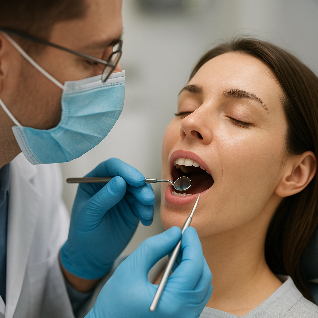
X-rays are a vital tool in modern dentistry, allowing practitioners to diagnose and treat various dental conditions effectively. However, the use of X-rays involves exposure to ionizing radiation, which can pose health risks if not managed properly. The latest guidelines emphasize the principle of ALARA (As Low As Reasonably Achievable), which aims to minimize radiation exposure while still obtaining the necessary diagnostic information.
The ALARA Principle
The ALARA principle is foundational in radiology and dentistry. It encourages dental professionals to take all reasonable steps to reduce radiation exposure to patients, staff, and themselves. This includes:
- Justification: Ensuring that every X-ray taken is necessary for diagnosis or treatment.
- Optimization: Using the lowest possible radiation dose to achieve the required image quality.
- Limitation: Limiting the number of X-rays taken and the area exposed to radiation.
By adhering to these principles, dental practitioners can significantly reduce the risks associated with X-ray exposure while still providing high-quality care.
Current Guidelines and Recommendations
The latest guidelines on X-ray safety in dentistry are issued by various organizations, including the American Dental Association (ADA) and the International Commission on Radiological Protection (ICRP). These guidelines cover several key areas:
- Patient Selection: Dentists should evaluate the need for X-rays based on the patient’s dental history, clinical examination, and risk factors.
- Radiation Dose Management: Use of digital radiography is encouraged, as it typically requires less radiation than traditional film-based methods.
- Protective Measures: The use of lead aprons and thyroid collars is recommended to protect patients from unnecessary radiation exposure.
- Quality Assurance Programs: Regular maintenance and calibration of X-ray equipment are essential to ensure optimal performance and safety.
These guidelines are designed to protect patients while ensuring that dental professionals can effectively diagnose and treat dental issues.
Advancements in Dental Imaging Technology
Technological advancements have played a significant role in enhancing X-ray safety in dentistry. Innovations in imaging techniques and equipment have led to reduced radiation doses and improved image quality. Some notable advancements include:
Digital Radiography
Digital radiography has revolutionized dental imaging by replacing traditional film with digital sensors. This technology offers several advantages:
- Reduced Radiation Exposure: Digital sensors are more sensitive to radiation, allowing for lower doses to achieve high-quality images.
- Immediate Image Availability: Digital images can be viewed instantly, reducing the need for retakes and further exposure.
- Enhanced Image Quality: Digital images can be manipulated for better visualization, aiding in accurate diagnosis.
As a result, digital radiography aligns well with the ALARA principle, making it a preferred choice in modern dental practices.
3D Imaging Techniques
Three-dimensional imaging techniques, such as Cone Beam Computed Tomography (CBCT), have also emerged as valuable tools in dentistry. CBCT provides detailed images of dental structures, allowing for more accurate diagnoses and treatment planning. While these techniques may involve higher radiation doses than traditional X-rays, the benefits often outweigh the risks when used judiciously.
Software Enhancements
Advancements in imaging software have further improved the safety and efficacy of dental X-rays. Features such as automatic exposure control and image processing algorithms help optimize radiation doses and enhance image quality. Additionally, software can assist in tracking patient exposure history, ensuring that practitioners adhere to safety guidelines.
Training and Education for Dental Professionals
To ensure compliance with the latest X-ray safety guidelines, ongoing training and education for dental professionals are essential. Dental schools and continuing education programs should emphasize the importance of radiation safety, the ALARA principle, and the latest technological advancements in imaging.
Continuing Education Programs
Many professional organizations offer continuing education courses focused on radiology and X-ray safety. These programs provide dental professionals with the knowledge and skills necessary to implement best practices in their practices. Topics may include:
- Radiation physics and biology
- Current guidelines and regulations
- Advancements in imaging technology
- Patient communication regarding X-ray safety
By participating in these programs, dental professionals can stay informed about the latest developments in X-ray safety and improve their practice’s overall quality of care.
Patient Education and Communication
Educating patients about X-ray safety is equally important. Dental professionals should take the time to explain the necessity of X-rays, the safety measures in place, and the steps taken to minimize radiation exposure. This transparency helps build trust and encourages patients to feel more comfortable with the imaging process.
Conclusion
The latest guidelines on X-ray safety in dentistry are vital for protecting patients and dental professionals alike. By adhering to the ALARA principle, utilizing advanced imaging technologies, and prioritizing ongoing education, dental practitioners can ensure that they provide safe and effective care. As technology continues to evolve, staying informed about best practices and safety measures will remain essential in the field of dentistry.