The use of X-rays has revolutionized the field of dentistry, offering clinicians an unparalleled window into the hidden structures of the oral cavity. These imaging techniques play a critical role in diagnosing conditions that cannot be detected through visual examination alone. By harnessing the power of radiation in controlled doses, dental professionals can reveal caries, infections, bone loss, and developmental anomalies, leading to more accurate treatment planning and better patient outcomes.
Fundamentals of Dental X-ray Imaging
Basic Principles
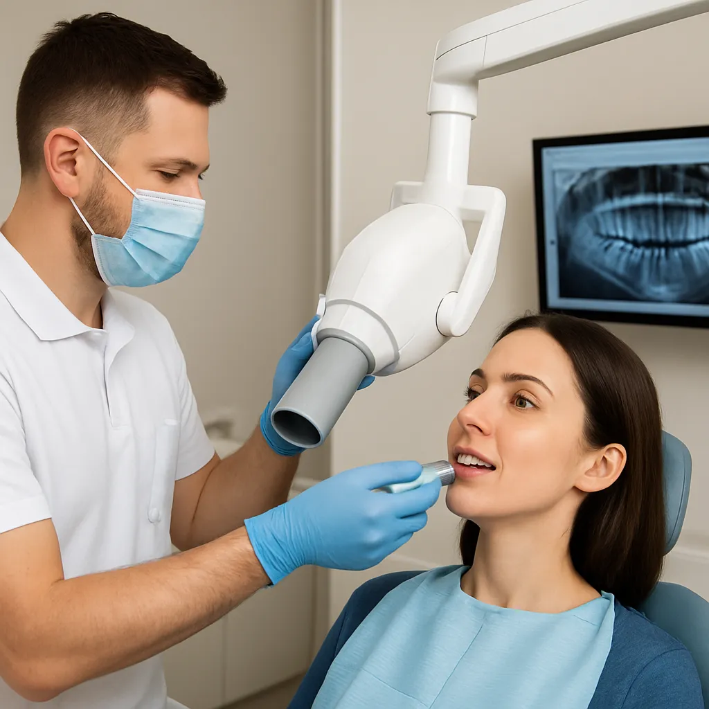
X-rays are a form of electromagnetic energy that can pass through soft tissues but are absorbed by denser materials like bone and teeth. The resulting image highlights these high-density areas, allowing dentists to identify pathologies invisible to the naked eye. The process involves generating a beam of X-ray photons, directing it toward the targeted area, and capturing the attenuated beam on a receptor, which produces a two-dimensional image.
Types of Dental Radiographs
- Periapical: Focuses on one or two teeth from root tip to crown, ideal for detecting abscesses or root fractures.
- Panoramic: Provides a broad view of the entire jaw, temporomandibular joints, and sinuses, useful for orthodontic assessment and implant planning.
- Bitewing: Captures the crowns of both upper and lower teeth in one image, excellent for detecting interproximal caries and monitoring bone levels.
- Cone Beam Computed Tomography (CBCT): Produces a three-dimensional reconstruction, enabling precise evaluation of bone volume and anatomical structures.
Clinical Applications in Modern Dentistry
Endodontics and Periapical Assessment
In endodontic therapy, accurate diagnosis of root canal anatomy and periapical health is essential. X-rays help determine the number of canals, identify unusual curvatures, and confirm the presence of periapical lesions. By using sequential radiographs, practitioners can evaluate the progress of root canal obturation and ensure complete debridement of infected tissues.
Orthodontics and Growth Prediction
Orthodontists rely on panoramic and lateral cephalometric radiographs to analyze skeletal relationships and tooth positioning. These images allow for assessment of growth patterns, detection of impacted teeth, and planning of complex tooth movements. Advanced digital imaging software can overlay treatment models, enhancing precision and reducing treatment time.
Implant Dentistry and Surgical Planning
Successful implant placement demands thorough preoperative evaluation of bone density, nerve location, and sinus anatomy. CBCT scans deliver high-resolution, three-dimensional data that guide the design of surgical guides and ensure implants are placed with optimal angulation and depth. This integration of imaging with guided surgery enhances the predictability of osseointegration and long-term success.
Safety and Radiation Protection
ALARA Principle
Adherence to the “As Low As Reasonably Achievable” (ALARA) principle is paramount in dental radiography. By selecting appropriate exposure settings, using fast imaging receptors, and employing beam-limiting devices such as collimators, practitioners can minimize patient dose without compromising diagnostic quality.
Protective Measures for Patients and Staff
- Lead aprons and thyroid collars: Shield sensitive organs from scatter radiation.
- Digital sensors: Require significantly less exposure compared to traditional film, reducing dose by up to 60–80%.
- Distance and barriers: Operators should maintain a safe distance or stand behind protective walls when activating the X-ray machine.
- Regular equipment maintenance: Ensures proper calibration and prevents dose creep over time.
Regulatory Guidelines and Quality Assurance
Compliance with national and international regulations, such as those promulgated by the International Atomic Energy Agency (IAEA) and local health authorities, is essential. Routine quality assurance tests, including beam alignment checks, output consistency assessments, and sensor integrity evaluations, guarantee that imaging systems perform within specified safety limits.
Advances in Digital Imaging and Interpretation
Integration with Electronic Records
Digital radiography seamlessly integrates with electronic health record systems, facilitating rapid image storage, retrieval, and sharing. Clinicians can annotate images, measure distances, and compare sequential radiographs, boosting diagnostic accuracy and patient communication.
Artificial Intelligence and Automated Analysis
Emerging technology in artificial intelligence (AI) promises to revolutionize the interpretation of dental X-rays. Algorithms trained on thousands of images can detect caries, periodontal bone loss, and periapical radiolucencies with high sensitivity and specificity. AI-driven decision support tools help practitioners identify subtle changes over time and prioritize cases requiring urgent intervention.
Three-Dimensional Visualization
CBCT and advanced reconstruction software enable clinicians to rotate and slice volumetric images, revealing the precise location of anatomical structures. This three-dimensional insight is invaluable for complex surgical procedures, such as sinus lifts, bone grafting, and nerve repositioning, reducing intraoperative surprises and enhancing patient safety.
Protocols for Effective Image Acquisition
Patient Positioning and Technique
- Stable head support: Ensures consistent positioning and minimizes motion artifacts.
- Proper receptor placement: Aligns the sensor parallel to the long axis of the tooth and perpendicular to the X-ray beam.
- Standardized exposure parameters: Tailored to patient size, age, and anatomical region.
- Use of film holders or positioning devices: Reduces retakes and improves image reproducibility.
Image Enhancement and Post-Processing
Digital systems offer tools for adjusting contrast, brightness, and sharpness. Filters can reduce noise or enhance edges, aiding in the identification of microfractures and early carious lesions. However, practitioners must apply enhancements judiciously to avoid introducing artifacts or obscuring critical details.
Future Directions and Research Trends
Low-Dose Imaging Modalities
Ongoing research focuses on developing new detector materials and X-ray generation techniques that further reduce radiation dose without sacrificing image quality. Innovations such as photon-counting detectors and ultrafast pulsed X-ray sources hold promise for safer, more detailed imaging.
Personalized Radiographic Protocols
The concept of personalized imaging protocols tailors exposure settings to individual patient characteristics, such as bone density and anatomical variations. By leveraging patient data and machine learning, future systems may automatically adjust parameters to deliver the optimal balance between dose and diagnostic value.
Integration with Other Diagnostic Modalities
Combining X-ray imaging with optical coherence tomography (OCT), near-infrared transillumination, and fluorescence-based caries detection can provide a more comprehensive assessment of tooth structure and pathology. Multimodal diagnostic platforms aim to detect early-stage lesions and monitor treatment progress with unprecedented precision.
Essential Considerations for Practice Implementation
Training and Continuing Education
Proficiency in dental radiography requires ongoing training to keep pace with technological advances and evolving safety standards. Certification programs and hands-on workshops help clinicians master image acquisition techniques, radiation protection protocols, and advanced interpretation skills.
Patient Communication and Informed Consent
Educating patients about the purpose, benefits, and minimal risks of X-ray examinations fosters trust and compliance. Clear explanations and visual aids can help alleviate concerns about safety and demonstrate the necessity of imaging for effective diagnosis and treatment.
Cost-Benefit Analysis
Investing in high-end imaging equipment involves significant financial outlay. Practices should conduct a thorough cost-benefit analysis, considering factors such as patient demand, case complexity, reimbursement policies, and potential improvements in treatment outcomes. Strategic planning ensures that technology investments align with practice goals and patient needs.