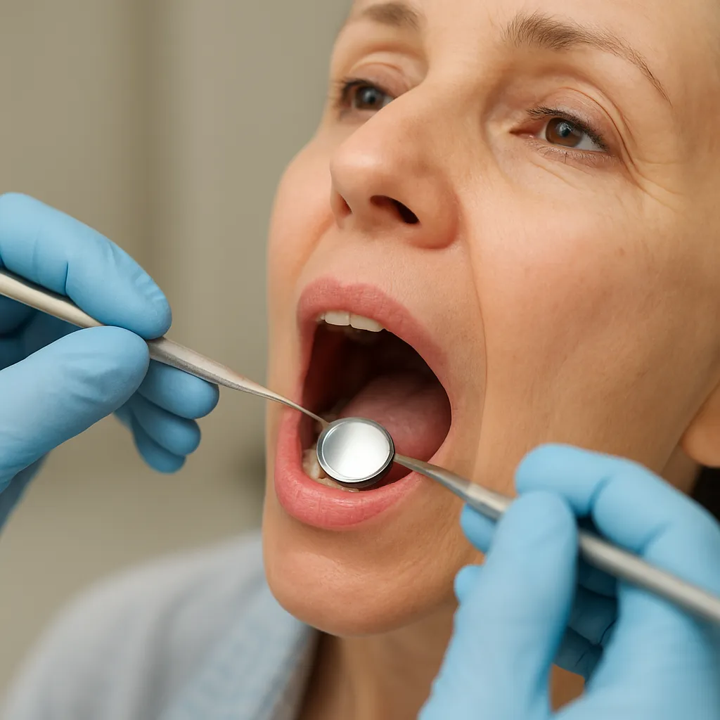
Oral cancer screening plays a vital role in the maintenance of a healthy mouth and overall well-being. As dental professionals, understanding each stage of the process ensures accurate examination and promotes patient confidence. A comprehensive screening helps identify abnormalities at the earliest possible moment, leading to better outcomes and increased survival rates. This article outlines key aspects of the screening journey, from initial risk assessment to appropriate referral when necessary.
Understanding Oral Cancer and Risk Factors
Detecting oral cancer in its early stages dramatically improves treatment success. Dentists should remain vigilant for any changes in the tissues of the lips, tongue, cheeks, and floor of the mouth. By becoming familiar with common risk factors, professionals can customize screening frequency and intensity for each patient.
What Is Oral Cancer?
Oral cancer involves malignant growths in the mouth’s soft tissues or salivary glands. It may present as persistent ulcers, red or white patches, or unexplained lumps. Because symptoms can mimic those of benign conditions, it’s crucial to conduct a thorough and systematic review of suspicious areas.
Main Risk Factors
- Tobacco use (cigarettes, cigars, pipes, chewing tobacco)
- Excessive alcohol consumption
- Human papillomavirus (HPV) infection
- Poor nutritional status and immune suppression
- Prolonged sun exposure (lip cancer risk)
Preparing for Your Oral Cancer Screening
Preparation begins long before your patient sits in the dental chair. Establish a trusting environment through clear communication, tailored questionnaires, and up-to-date medical histories. Encourage patients to report any discomfort, unexplained bleeding, or changes in sensation.
Reviewing Medical History
A detailed history uncovers predispositions to malignancy. Ask about previous cancer diagnoses, family history, tobacco and alcohol use, and any recent weight loss or fatigue. Be attentive to immune-compromised conditions, such as HIV or organ transplant status, which can increase cancer risk.
Communicating Symptoms
Patients often hesitate to mention minor discomfort or persistent soreness. Use open-ended questions to elicit relevant details: when did the lesion appear, has it changed size or color, and is there pain on swallowing? Document every symptom accurately to inform your clinical decision-making.
Step-by-Step Oral Cancer Examination
A systematic approach ensures no area is overlooked. Incorporate both visual and tactile methods to evaluate the entire oral cavity and nearby structures. Consistency and thoroughness are key to high-quality screenings.
Visual Inspection
Under optimal lighting, inspect the lips, vestibule, buccal mucosa, gingiva, tongue (dorsal, ventral, lateral), floor of the mouth, palate, and oropharynx. Look for color changes (lesion presence), texture alterations, or irregular growths. Document any red (erythroplakia) or white (leukoplakia) areas immediately.
Palpation Techniques
Use gloved fingers to gently palpate the floor of the mouth and external lymph node regions (submandibular, cervical). Evaluate for firmness, nodularity, or fixation to underlying tissues. Palpation helps assess the depth and extent of any unexpected masses.
Use of Adjunctive Tools
Adjunctive devices, such as VELscope or toluidine blue staining, can highlight suspicious areas invisible to the naked eye. While these tools offer additional insight, they should complement—not replace—a skilled clinical examination.
Dealing with Suspicious Findings
When anomalies are detected, swift action is essential. Prompt biopsy and histopathological analysis provide definitive diagnoses. Early intervention can significantly reduce the risk of metastasis and improve prognosis.
Lesion Documentation
Photographic records and accurate measurements assist in monitoring lesion progression. Capture high-resolution images from multiple angles and record precise dimensions. A well-documented case history supports future comparisons and legal defense, if necessary.
Biopsy and Referral
Depending on lesion characteristics, choose an incisional or excisional biopsy. If surgical removal falls outside your practice’s scope, arrange a timely referral to an oral surgeon or head and neck specialist. Clear communication with the specialist—including documentation of your findings—ensures continuity of care.
Best Practices and Professionalism
Maintaining a professional yet empathetic demeanor fosters patient trust. Emphasize the importance of regular screenings and lifestyle modifications. By integrating cancer checks into every routine examination, dental teams demonstrate a commitment to comprehensive care.
Educating the Patient
Discuss preventive strategies such as tobacco cessation, reduced alcohol intake, and safe sexual practices to lower HPV risk. Encourage patients to perform self-examinations monthly, checking for any new or persistent abnormalities.
Follow-Up Scheduling
Set clear intervals for reevaluation based on initial findings and risk profile. Patients with suspicious but non-conclusive results might need closer surveillance—often every three months—whereas low-risk patients may return for annual checks. Robust follow-up protocols can catch changes before they escalate.