Many patients experience persistent discomfort in their upper teeth without obvious dental problems. This pain often has a source beyond the oral cavity. Sinus infections can cause referred dental pain, making accurate diagnosis essential. Understanding the intricate relationship between the maxillary sinuses and upper molars helps clinicians differentiate between typical toothaches and those originating from sinus inflammation.
Anatomy of the Oral and Sinus Regions
Accurate knowledge of the anatomical structures is crucial for both dentists and ENT specialists. The close proximity of the teeth roots to the sinus floor creates a potential pathway for inflammatory processes to affect dental tissues. Discerning these connections minimizes unnecessary procedures like extraction or root canal therapy when the problem stems outside the tooth.
Maxillary Sinuses and Upper Teeth
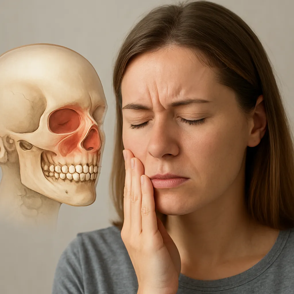
The paired sinus cavities located above the upper jaw are called the maxillary sinuses. Each cavity lies directly above the premolars and molars. In many individuals, the roots of the second molars may even protrude into the sinus floor. When these sinuses become infected or congested, they can press on tooth roots, causing tooth pain that mimics genuine dental pathology.
- Sinus floor proximity: thin bone or dehiscence allows pressure transmission.
- Root anatomy variations: long roots may intrude the sinus space.
- Mucosal lining: inflammation can increase pressure and fluid build-up.
Neurological Convergence
The trigeminal nerve supplies both the teeth and sinus mucosa. This shared innervation leads to referred pain, where sinus discomfort is perceived as a dental ache. For instance, branches of the maxillary division (V2) convey signals from the upper teeth and sinus membranes. Irritation of these fibers triggers similar pain pathways.
How Sinus Infections Lead to Tooth Pain
Typical sinus infections, or sinusitis, arise from viral or bacterial agents. The resulting congestion and fluid retention increase sinus pressure. This buildup translates into an unpleasant sensation in the maxillary region, which patients often interpret as coming from their back teeth.
Mechanism of Referred Pain
- Inflammation of sinus mucosa leads to swelling.
- Increased intraluminal pressure presses on adjacent roots.
- Stimulation of nociceptors within the trigeminal nerve branches.
- Perception of pain localized at the tooth apexes.
Clinical Presentation
Sinus-related dental pain typically:
- Worsens when bending forward or lying down due to fluid shift.
- Coexists with nasal congestion, discharge, and facial pressure.
- May involve several adjacent teeth rather than a single focal point.
- Does not always respond to conventional cavity treatments.
In contrast, true dental pain often:
- Is sharp and localized to one tooth.
- Aggravates with hot or cold stimuli.
- Intensifies upon biting or tapping the tooth surface.
Diagnosis and Management Strategies
Effective management begins with distinguishing sinus origin from dental origin. A collaborative approach between dentists and ENT experts ensures accurate diagnosis and avoids unnecessary interventions.
Clinical Examination
- Thorough history: Document onset, duration, aggravating factors.
- Palpation: Assess tenderness over the malar region.
- Percussion tests: Evaluate tooth response to tapping.
- Transillumination: Helps detect sinus fluid accumulation.
Imaging Techniques
Radiographic evaluation plays a pivotal role. Dentists rely on periapical and panoramic radiographs, but these may not reveal sinus pathology. Collaboration with ENT allows advanced imaging:
- CT scan: identifies mucosal thickening, fluid levels, and anatomical variants.
- MRI: differentiates between soft tissue changes and neoplastic processes.
- Ultrasound: a non-invasive tool for superficial sinus assessment.
Microbiological Assessment
In chronic cases, obtaining sinus aspirate for culture guides targeted antibiotic therapy. This step differentiates viral from bacterial sinusitis, preventing overuse of broad-spectrum agents.
Treatment Options
Once sinus origin is confirmed, dental procedures can be deferred unless dental pathology coexists. Treatment includes:
- Antibiotics: Amoxicillin-clavulanate or doxycycline for bacterial infections.
- Decongestants: Intranasal sprays or oral pseudoephedrine.
- Corticosteroid nasal sprays to reduce mucosal inflammation.
- Saline irrigation for mechanical clearance of secretions.
- Analgesics and pain management protocols for symptomatic relief.
When to Refer to a Specialist
Persistent or complicated sinusitis may require an ENT consultation. Indications include:
- Failure of medical therapy after 10–14 days.
- Recurring infections (≥3 episodes per year).
- Evidence of anatomical obstruction on imaging.
- Signs of orbital or intracranial spread.
Preventive Measures and Patient Education
Preventing recurrent sinusitis-related tooth pain involves both general and dental-specific strategies. Patients benefit from understanding how lifestyle and oral hygiene affect their sinuses.
- Maintain good nasal hygiene with saline rinses.
- Manage allergies with appropriate medications.
- Avoid tobacco smoke and known sinus irritants.
- Stay well-hydrated to thin mucus secretions.
- Regular dental check-ups to preempt odontogenic infections.
Educating patients about the signs of sinus versus dental pain empowers them to seek timely care and reduces unnecessary dental treatments. When clinicians use a structured approach and clear communication, outcomes improve and patient satisfaction rises.