Removing wisdom teeth is one of the most common oral surgical procedures performed by dentists and oral surgeons. These third molars often erupt during late adolescence or early adulthood, and their arrival can present various challenges. Understanding when removal is recommended, what to expect during the procedure, and how to manage healing can help patients feel more confident and prepared. This article explores key aspects of wisdom teeth removal, from initial consultation to full recovery, providing dental professionals and patients with practical insights.
What Are Wisdom Teeth and Why They Matter
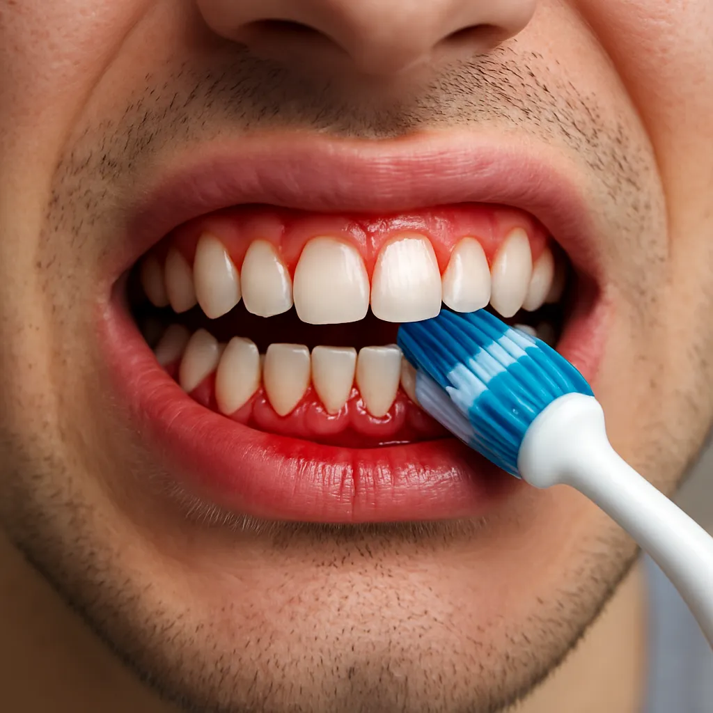
Wisdom teeth, also called third molars, typically emerge between ages 17 and 25. While some patients develop sufficient jaw space to accommodate these extra molars, many experience complications because these teeth become impacted or grow at awkward angles. Impacted wisdom teeth can press against neighboring molars, leading to crowding, pain, and enamel damage. In other cases, partially erupted teeth create a pocket that harbors bacteria, increasing the risk of gum disease and infection.
Clinicians evaluate radiographs to determine the position and health of third molars. Panoramic X-rays and 3D cone-beam computed tomography (CBCT) scans reveal root structure, proximity to the mandibular nerve, and bone density. Early assessment allows dental professionals to decide whether to monitor growth or recommend timely removal, potentially reducing the severity of complications.
When Should Wisdom Teeth Be Removed?
Deciding on extraction involves balancing potential benefits and risks. Many practitioners follow these guidelines:
- Asymptomatic but impacted teeth that threaten adjacent molars.
- Partially erupted molars with soft tissue inflammation or recurring pericoronitis.
- Evidence of cyst formation, root resorption, or signs of decay.
- Orthodontic treatment plans requiring clear posterior alignment.
In certain cases, prudent monitoring with periodic radiographs may suffice. However, extraction is often recommended before the roots fully develop and the bone becomes denser. This approach typically leads to faster healing and fewer post-operative complications.
The Extraction Process: Step by Step
Consultation and Preparation
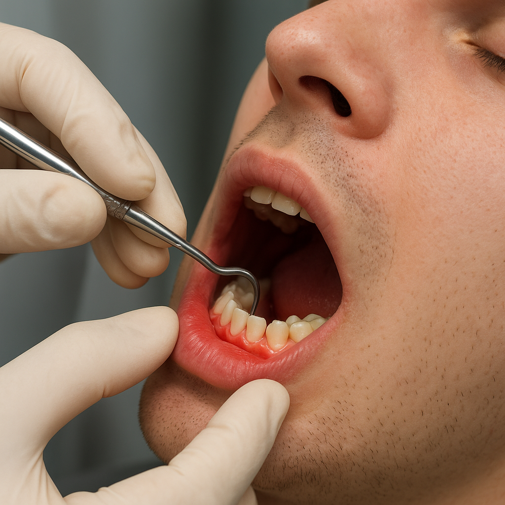
During the initial visit, the dentist reviews the patient’s medical history, discusses any allergies, and addresses concerns about anesthesia options—local, intravenous sedation, or general. Dental professionals explain risks, benefits, and alternatives, ensuring informed consent.
Surgical Procedure
On the day of surgery, the clinician administers the chosen anesthesia. For complex extractions, IV sedation or general anesthesia may be preferred. Once the patient is comfortable:
- An incision is made in the gum tissue to access the tooth and bone.
- A small amount of bone may be removed to expose the tooth crown.
- The tooth is sectioned in cases of multi-rooted molars to facilitate removal.
- After extraction, the socket is cleaned and irrigated to remove debris.
- If bone loss or structural support is a concern, a bone graft may be placed to preserve the alveolar ridge.
- Sutures are applied to promote healing and protect the surgical site.
Throughout the procedure, gentle tissue handling, adequate visualization, and hemostasis are critical to minimize trauma and postoperative discomfort.
Post-Operative Care and Recovery
Proper aftercare significantly affects healing outcomes. Patients should follow these recommendations:
- Avoid spitting or using straws for the first 24 hours to protect the blood clot.
- Apply an ice pack intermittently to reduce swelling.
- Maintain a soft diet—yogurt, mashed potatoes, smoothies—for at least 48–72 hours.
- Brush gently around the surgical site, keeping sutures and adjacent teeth clean.
- Take prescribed pain relievers and antibiotics as directed to prevent infection.
- Refrain from smoking and vigorous exercise for a week to aid clot stability.
Regular follow-up visits allow dentists to assess healing, remove sutures if necessary, and address any patient concerns. Complete recovery generally occurs within two weeks, though minor discomfort can persist for a short time.
Potential Complications and Prevention
While most extractions proceed without major issues, practitioners must remain vigilant for complications:
- Dry Socket: Occurs when the clot dislodges, exposing bone and nerve endings, leading to severe pain. Prevention involves careful post-op instructions and avoiding dislodgement.
- Nerve Injury: Inferior alveolar or lingual nerve damage can cause temporary or, rarely, permanent numbness. Preoperative imaging helps map nerve pathways.
- Excessive Bleeding: Patients on anticoagulants require tailored protocols to manage bleeding risk.
- Alveolar Osteitis: Inflammation of the bone can result in prolonged recovery; maintaining proper hygiene and hydration mitigates this risk.
By educating patients on warning signs—persistent bleeding, escalating pain, or unusual swelling—clinicians ensure prompt management. Should complications arise, timely intervention with irrigation, medicated dressings, or secondary procedures promotes resolution.