Regular dental X-rays play a crucial role in maintaining optimal oral health by enabling dentists to visualize hidden structures and detect issues before they manifest as pain or visible problems. These radiographic images provide invaluable insights into the condition of teeth, gums, and supporting bone, empowering both patients and professionals to take proactive steps toward comprehensive care.
Benefits of Regular Dental X-rays
- Detection of early decay: Subtle cavities between teeth or beneath existing fillings often remain invisible during a clinical exam. Radiographs reveal these hidden issues, allowing for prompt intervention.
- Identification of bone loss: Periodontal disease erodes the jawbone over time. X-rays highlight areas of bone deterioration, guiding targeted periodontal therapy.
- Evaluation of unerupted teeth: Wisdom teeth and impacted canines can be monitored through X-rays to determine their orientation and potential complications.
- Assessment of developmental anomalies: Congenital or acquired defects in tooth shape, number, or structure become clear on radiographs, aiding in appropriate orthodontic or restorative planning.
- Monitoring of existing restorations: Regular imaging helps evaluate the integrity of crowns, bridges, and implants, spotting any recurrent decay or mechanical failure at an early stage.
Early Intervention and Prevention
By discovering minor issues before they escalate, dentists can prescribe minimally invasive procedures such as fluoride treatments, sealants, or conservative restorations. This approach saves time, reduces costs, and minimizes patient discomfort.
Enhanced Treatment Planning
Comprehensive treatment plans rely on accurate information. X-rays inform decisions on root canal therapy, implant placement, and orthodontic movements by visualizing root morphology, bone density, and spatial relationships.
Common Types of Dental X-rays
Different radiographic techniques serve specific diagnostic purposes. Understanding the variety of imaging methods helps patients appreciate the versatility and value of each procedure.
- Intraoral Radiographs
- Periapical X-rays: Capture entire tooth structures from crown to root, ideal for detecting root infections and evaluating periodontal bone.
- Bitewing X-rays: Focus on the crowns of posterior teeth, excellent for uncovering interproximal decay and assessing marginal bone levels.
- Occlusal X-rays: Provide a broad view of one arch, useful for locating supernumerary teeth, fractures, or cysts in young children.
- Extraoral Radiographs
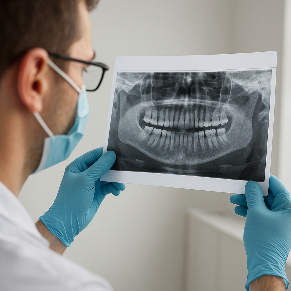
- Panoramic X-rays: Offer a wide overview of both jaws, sinuses, and temporomandibular joints, advantageous for surgical planning and growth assessment.
- Cephalometric Radiographs: Record lateral skull images for orthodontic analysis of jaw relationships and facial growth.
- 3D Imaging (CBCT)
Cone-beam computed tomography generates three-dimensional images, delivering precise details on bone volume and anatomical landmarks. CBCT is indispensable for complex implant placement and evaluation of maxillofacial pathology.
Addressing Safety and Frequency Concerns
Although X-rays involve exposure to ionizing radiation, modern dental practices adhere to strict protocols that minimize risk. Digital sensors require significantly less radiation than traditional film, and protective measures further reduce patient dose.
ALARA Principle
The acronym “ALARA” stands for “As Low As Reasonably Achievable.” Dentists balance diagnostic benefit against radiation exposure by:
- Selecting the appropriate imaging modality for each clinical scenario.
- Employing lead aprons and thyroid collars for additional protection.
- Using digital radiography to cut exposure levels by up to 90% compared to analog film techniques.
Recommended X-ray Intervals
Individual patient factors—such as age, risk of caries, and periodontal health—dictate imaging schedules. As a general guideline:
- New patients often receive a full-mouth series upon initial evaluation to establish a baseline.
- Recall X-rays for low-risk adults may be suggested every 18–24 months, focusing on bitewings.
- High-risk individuals, children, and those undergoing active treatment may require imaging every 6–12 months.
Integrating Dental X-rays into Comprehensive Care
Effective oral healthcare combines clinical examination with radiographic analysis to form a clear, complete picture of a patient’s status. This integrated approach ensures that underlying pathology does not go unnoticed.
Collaboration and Referral
When radiographic findings suggest complex conditions—such as cysts, tumors, or severe bone defects—general dentists collaborate with specialists, including oral surgeons and endodontists, for advanced management.
Patient Education and Engagement
Sharing radiographic images during consultations fosters patient understanding and encourages active participation in treatment decisions. Visual evidence of decay or bone changes reinforces preventive recommendations and underscores the importance of regular visits.
Future Trends in Dental Imaging
Innovations continue to enhance diagnostic capabilities and patient comfort. Emerging technologies include:
- 3D printing integration with CBCT data, allowing fabrication of surgical guides and custom prosthetics.
- Artificial intelligence algorithms that analyze radiographs to identify subtle changes, offering second-opinion support and boosting diagnostic accuracy.
- Portable imaging units that facilitate on-site radiography in community outreach programs, expanding access to underserved populations.
Emphasis on Minimally Invasive Dentistry
Advancements in technology aim to further reduce radiation exposure and improve image resolution. As equipment evolves, dentists will detect lesions at even earlier stages, aligning with the philosophy of minimally invasive dentistry.
Conclusion of Key Points
Regular dental X-rays represent a cornerstone of preventive and restorative care. By harnessing the power of radiographic imaging, clinicians achieve accurate diagnosis, tailor treatment planning, and safeguard long-term oral function. Patients benefit from early detection of issues, less invasive interventions, and sustained oral health. Embracing the advantages of modern radiography within a framework of stringent safety measures ensures that both practitioners and patients reap maximum rewards from this indispensable tool.